JUNIOR INTER ZOOLOGY MOST EXPECTED QUETIONS WITH ANSWERS
| zoology-examseries-1.pdf |
1. Describe the digestive system in Pheretima with the help of a diagram?
(March 2005, May 2007, March 2010 , MARCH 2011)
Ans: The digestive system consists of the alimentary canal and the associated digestive
glands. The alimentary canal consists of the following parts.
1) MOUTH
* It is a crescentic aperture present at the anterior end, ventrally on the first segment called Peristomium
* The extension of the first segment over hangs like a lip infront of the mouth, called prostomium.
2) BUCCAL CHAMBER
* The mouth leads into Buccal chamber.
* The Buccal chamber extends from the first to the middle of the third segments
* The walls of the Buccal chamber turn inside out and the Pharynx ( everts ) comes out through the mouth
and helps in digging the soil.
3) PHARYNX
* Buccal chamber leads into Pharynx.
* It occurs in the third and fourth segments.
* Its wall is muscular and contains several blood cells.
* Its Dorsal side is folded and consists of a glandular structure called Pharyngeal bulb, thelateral wall is
pushed inwards to form a pair of lateral folds called shelves.
* They divide the pharyngeal cavity into 2 compartments, called the dorsal salivary chamber and the ventral
conducting chamber.
* The chromophilic cells of Pharyngeal bulb secrete proteolytic enzymes and mucus that help in protein digestion.
* The ventral conducting chamber helps in passing the food.
4) OESOPHAGUS
* The Pharynx opens into the oesophagus.
* The oesophagus is a narrow tube extending from the 5th to 8th segments.
5) GIZZARD
* Oesophagus in 8th segment opens into the gizzzard .
* It is an oval shaped hard muscular chamber .
* It has thick wall of circular muscles and inner lining of chitinous cuticle.
* It grinds the food material into fine particles.
* It is commonly called “ grinding mill “
6) STOMACH
* The gizzard leads into the stomach.
* The stomach extends from the 9th to the 14th segment.
* It is a highly vascular and glandular tube.
* Its inner foldings contain gland cells, which secrete proteolytic enzymes, that help in protein dig estion.
7) INTESTINE :
* Stomach opens into the long and wide intestine which extends from the 15th
to the last segment.
* It’s mid dorsal fold is called typhlosole. This is poorly developed in Pheretima.
* It increases the area of absorption of food.
* On the basis of the presence of typhlosole the intestine is divisible into 3 regions.
1) Pre-typhlosolar region 2) Typhlosolar region 3) Post-typhlosolar region
1) Pre-typhlosolar region :
* This is the proximal part of the intestine located from 15th
to 25th segments.
* In the 26th segment, a pair of conical outgrowths, the intestinal caecae arise form this region laterally
and extend forward over three or four segments.
* They secrete digestive juices, which contain amylase enzyme.
* Typhlosole is absent in this part of intestine.
2) Typhlosolar region :
* It starts from 26th segment and terminates in 23 or 25 segments ahead of the hind end of the body.
* It is distinguished by the presence of small mid dorsal projection, the typhlosole, hanging into the
lumen of the intestine. Typhlosole is poorly developed in Pheretima.
* Digested food is absorbed in this region.
3) Post-typhlosolar region :
* It is the distal part of the intestine and it occupies the last 23 or 25 segments.
* It is without a typhlosole.
* It is also called rectum.
* The undigested food is converted into small pellets.
8) ANUS
* It is present in the last segment. The undigested food is sent out through the anus in the form of
pellets. These pellets are called “worm castings”.
PHERETIMA - IMP QUESTIONS
| pheretima.pdf |
IMP QUESTIONS FOR IPE
1. Define biotechnology and write its applications
Ans : It is the branch of biology that deals with the use of biological agents,such as microorganisms and certain cellular components,for beneficial purposes.
Applications of biotechnology :-
a) Pollution control
b) Pharmacology
c) Production of transgenic animals
d) Public heath
2. Distinguish between Eugenics and Euphenics
Ans: EUGENICS:- It is the branch of genetics that deals with the application of knowledge of genetics to human welfare.
EUPHENICS :- It is brach of genetics that deals with the practice of phenotypic improvement of humans after birth.
3. What are retroperitoneal organs ?
Ans : The organs of vertebrates that occur outside the coelom and are covered by peritoneum only on the surface facing the coelom are called “retroperitoneal organs”
4. List out the four types of cells present in the epidermis of Pheretima
Ans : The four types of cells in the epidermis of Pheretima are :
1) large glad cells 2) Supporting cells 3) Basal cells 4 ) Receptor cells
FLAGELLA AND CILIA
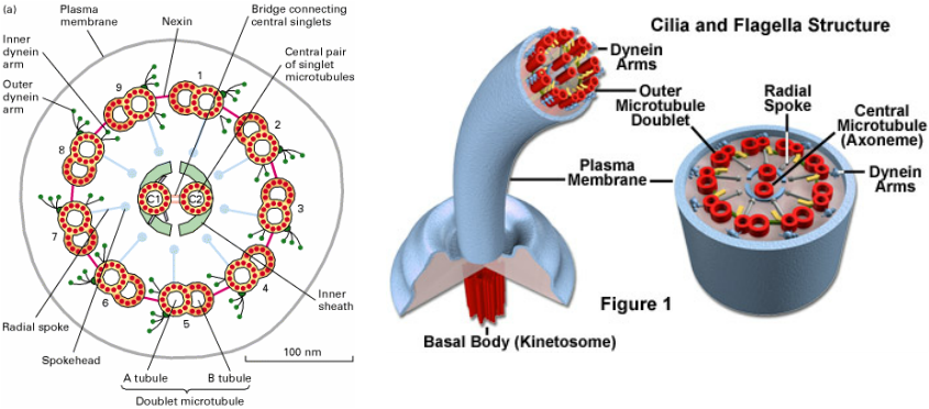
Cilia and flagella are motile cellular appendages found in most microorganisms and animals, but not in higher plants. In multicellular organisms, cilia function to move a cell or group of cells or to help transport fluid or materials past them. The respiratory tract in humans is lined with cilia that keep inhaled dust, smog, and potentially harmful microorganisms from entering the lungs. Among other tasks, cilia also generate water currents to carry food and oxygen past the gills of clams and transport food through the digestive systems of snails. Flagella are found primarily on gametes, but create the water currents necessary for respiration and circulation in sponges and coelenterates as well. For single-celled eukaryotes, cilia and flagella are essential for the locomotion of individual organisms. Protozoans belonging to the phylum Ciliophora are covered with cilia, while flagella are a characteristic of the protozoan group Mastigophora.
In eukaryotic cells, cilia and flagella contain the motor protein dynein and microtubules, which are composed of linear polymers of globular proteins called tubulin. The core of each of the structures is termed the axoneme and contains two central microtubules that are surrounded by an outer ring of nine doublet microtubules. One full microtubule and one partial microtubule, the latter of which shares a tubule wall with the other microtubule, comprise each doublet microtubule (see Figure 1). Dynein molecules are located around the circumference of the axoneme at regular intervals along its length where they bridge the gaps between adjacent microtubule doublets.
A plasma membrane surrounds the entire axoneme complex, which is attached to the cell at a structure termed the basal body (also known as a kinetosome). Basal bodies maintain the basic outer ring structure of the axoneme, but each of the nine sets of circumferential filaments is composed of three microtubules, rather than a doublet of microtubules. Thus, the basal body is structurally identical to the centrioles that are found in the centrosome located near the nucleus of the cell. In some organisms, such as the unicellular Chlamydomonas, basal bodies are locationally and functionally altered into centrioles and their flagella resorbed before cell division.
Eukaryotic cilia and flagella are generally differentiated based on size and number: cilia are usually shorter and occur together in much greater numbers than flagella, which are often solitary. The structures also exhibit somewhat different types of motion, though in both cases movement is generated by the activation of dynein and the resultant bending of the axoneme. The movement of cilia is often described as whip-like, or compared to the breast stroke in swimming. Adjacent cilia move almost simultaneously (but not quite), so that in groups of cilia, wave-like patterns of motion occur. Flagella, however, exhibit a smooth, independent undulatory type of movement in eukaryotes. Prokaryotic flagella, which have a completely different structure built from the protein flagellin, move in a rotating fashion powered by the basal motor.
Defects in the cilia and flagella of human cells are associated with some notable medical problems. For example, a hereditary condition known as Kartagener's syndrome is caused by problems with the dynein arms that extend between the microtubules present in the axoneme, and is characterized by recurrent respiratory infections related to the inability of cilia in the respiratory tract to clear away bacteria or other materials. The disease also results in male sterility due to the inability of sperm cells to propel themselves via flagella. Damage to respiratory cilia may also be acquired rather than inherited and is most commonly linked to smoking cigarettes. Bronchitis, for instance, is often triggered by a build-up of mucus and tar in the lungs that cannot be properly removed due to smoking-related impairment of cilia.
JUNIOR INTER ZOOLOGY GUESS PAPER MARCH-2011
| jr-zoology-guess-paper-march-2011.pdf |
| wuchereria-lifecycle.pdf |
VERY IMPORTANT QUESTIONS AND ANSWERS FOR IPE
LAQs
1.Describe the human phase of Plasmodium vivax
LIFECYCLE OF PLASMODIUM IN MAN
PLASMODIUM LIFECYCLE IN MAN ;1.Plasmodium completes its life cycle in two hosts
2.female anopheles mosquito is definitive host , man is intermediate host
3.In man asexual lifecycle occurs
4.In man plasmodium completes its lifecycle in two stages. They are
a)Exoerythrocytic stage b) Erythrocytic stage
EXOERYTHROCYTIC STAGE: In this stage two generations are formed. They are preerythrocytic generations and exoerythrocytic generations
PREERYTHROCYTIC GENERATIONS :
1.When mosquito bite humanbeing the sporozoite stage of plasmodium enters into man
2.The sporozoite stage is infective stage of plasmodium to man
3.Sporozoites enters into liver cells from blood stream
4.After entering into the liver cells sporozoites are transformed into trophozoites in livercells
5.The nucleus of the trophozoite undergoes multiplications to become schizont
6.Within 8 days,cytoplasm divides and large number of cryptozoites are released into the liver sinusoids
7.These cryptozoites may either enter into RBC or into another livercells.
EXOERYTHROCYTIC GENERATIONS:
1.When the cryptozoites enter into livercells ,they transform into trophozoites in livercells
2.The nucleus of the trophozoite divides several times and become a multinucleate schizont
3.Then cytoplasm divides and large number of metacryptozoites are released into the sinusoids of liver
4.Two types of metacryptozoits are present ,they are a)Macrometacryptozoites and b)Micrometacryptozoites.
5.Macrometacryptomerozoite enter into fresh liver cells and micrometacryptozoites into RBC
ERYTHROCYTIC STAGE (GOLGI CYCLE) :
1.Micrometacryptozoites or cryptozoites enter into RBC and become trophozoites
2.The vacuole of the trophozoite increases in size and pushes the cytoplasm and nucleus to periphery
3.In this stage the parasite appears like a ring hence it is called signet ring stage
4.After signet ring stage plasmodium develops pseudopodia and appears like amoeba , hence this stage is called amoeboid stage
5.Later amoeboid stage develops into schizont
6.Again in this schizont multiplications take place and merozoites are formed
7.Merozoites are released from RBC along with haemozoin granules.
8.Due to this haemozoin granules the symptoms of malaria are seen
2. Describe the mosquito of Plasmodium vivax.
PLASMODIUM-MOSQUITO PHASE
1.After repeated erythrocytic lifecycles in man the merozoite become inactive and transformed into gametocytes.
2.There are two types of gametocytes ,a)Macrogametocytes
b)Microgametocytes
3.Macrogametocytes are called female gametocytes and Microgametocytes are called male gametocytes.
4.The further development of the gametocytes needs female anopheles mosquito.
5.When the female anopheles mosquito bites a malaria patient the male and female gametocytes enter into the crop of mosquito
6.Male gametocytes develop into male gametes and female gametocytes into female gametes.
7.The male and female gametes are fused to form a zygote
8.Intially the zygote is stationary later it becomes motile
9.The motile zygote is called ookinete
10.The ookinete enters into the crop wall of mosquito
11. In the crop wall it gets encysted
12.The encysted zygote is called oocyst
13.In oocyst it enlarges and undergoes multiplications
14.Due to multiplications sporoblasts are formed
15.The cyst containing sporoblasts is called sporocysts
16.Sporoblasts develop into sporozoites
17.Sporozoites released from sporocyst and reach salivary glands
18.When this mosquito with sporozoites in its salivary glands bites a man ,the sporozoites enter into man and life cycle stars in man
3.Describe the life cycle of Taenia sodium.
TAENIA SOLIUM LIFE CYCLE
LIFECYCLE OF TAENIA IN HUMAN :
1.Fertilization takes place in ootype
2.After fertilization zygote is formed
3.Zygote ,vitelline cell and vitelline shell are together called capsule
4.Many capsules roll into uterus
5.Rest of the embryonic development takes place in the uterus
6.Zygote is divided into two unequal cells called megamere and embryonic cell
7.Embryoinc cell divides into mesomeres and micromeres
8.Mesomeres develop into inner embryonic membrane
9.Megameres develop into outer embryonic membrane
10.Micromeres form hexacanth
11.Inner embryonic membrane ,outerembryonic membrane and hexacanth are together called hexacanth larva or oncosphere
12.Gravidproglatids containing oncospheres pass out through faeces
13.Oncosphere is the infective stage to pig
LIFECYCLE OF TAENIA IN PIG;
1.When pig consumes the faeces containing gravidproglatids ,oncospheres in gravid proglatids enter into the stomach of pig
2.The outer and inner embryonic membranes are dissolved in the intestine and hexacanth larvae are released
3.Hexacanth larvae are attached to the wall of the intestine
4.Hexacanth larvae penetrate the intestine wall and reach the liver
5.Hexacanth larvae reach the heart from the liver
6.From heart hexacanth reach the skeletal muscles of pig
7.In skeletal muscles hexacanth loses its hooks and develop into cysticercus cellulosae.
8.The pork containing cysticercus cellulosae is called measly pork
9.When man consumes measly pork cysticercus enters into man and develops into an adult
10.The infective stage of taenia to man is cycticercus cellulose
4.Describe the digestive system in Pheretima with the help of diagram
ANS: DIGESTIVE SYSTEM;
Alimentary canal and associated digestive glands are collectively called digestive system.
ALIMENTARY CANAL ; It consists of Mouth , buccal cavity, pharynx , oesophagus,gizzard,stomach,intestine .
Mouth :-It is located in the first segment. Due to the presence of the mouth the first segment is called “peristomium”. It leads into buccal cavity.
Buccal cavity :-It extends up to the middle of the yhird segment.It leads into Pharynx.
Pharynx :-It extends between middle of the third segment and end of the fourth segment.it has pharyngeal bulb which secretes proteolytic enzyme.Pharynx leads into oesophagus.
Oesophagus :-It extends between fifth segment to eighth segment.
Gizzard :- Oesophagus is modified into gizzard in eighth segment. It is muscular and grinds the food, hence it is called grinding mill.It leads into stomach.
Stomach :- It extends between ninth and fourteenth segment . It also secretes proteolytic enzymes.It leads into intestine.
Intestine :-It extends between fifteenth segment and last segment. It has three parts :a)Pre-typhlosolar region b) Typhlosolar region c)Post-typhlosolar region.
a)Pre-typhlosolar region:- Typhlosole is absent
b) Typhlosolar region:- Typhlosole is present
c)Post-typhlosolar region;- Typhlosole is absent , it is also called rectum. It opens to the out side through anus.
Anus :- It is present in the last segment.
5.Explain the blood circulation in the first thirteen segments in Phertima with a suitable diagram
ANS:- CIRCULATORY SYSTEM IN FIRST 13 SEGMENTS:-
BLOOD CIRCULATORY SYSTEM IN THE FIRST 13 SEGMENTS OF PHERETIMA
ARRENGEMENT OF BLOOD VESSELS IN THE FIRST 13 SEGMENTS OF PHERETIMA:-
In the first 13 segments of Pheretima the following bloodvessels are present
I)Longitudinal blood vessels II) The hearts
III)The anterior loops IV)Ring vessels
I) Longitudinal blood vessels:
There are 5 longitudinal trunks in Pheretima in its first 13 segments,they are :
1.One Dorsal blood vessel 2.One Ventral blood vessel
3.Two lateral oesophageal blood vessels
4.One supra oesophageal blood vessel
1. Dorsal blood vessel:
a.It is the largest blood vessel
b.It is muscular
c.It is contractile
d.It is valvular
e.Blood flows from posterior to anterior
f.It acts as a distributing blood vessel in the first 13 segments
g.It distributes blood to parts of alimentary canal from buccal cavity to stomach and pharyngeal nephridia
2.Ventral blood vessel:-
a.It is the longest blood vessel
b.It is nonmuscular
c.It is noncontractile
d.It is nonvalvular
e.Blood flows from anterior to posterior
f.It is a chief distributing blood vessel
g.It distributes blood to bodywall,septa,integumentary nephridia,repeoductive organs in the first 13 segments
3. Lateral oesophageal blood vessels:
a.One pair of lateral oesophageal blood vessels present either side of alimentary canal in the first 13 segments
b.These blood vessels collect blood from bodywall,septa,integumentary nephridia,repeoductive organs in the first 13 segments
4. SUPRA OESOPHAGEAL BLOOD VESSEL:-
a.It is located between 9 th segment to 13 th segment
b.It collects blood from gizzard ,stomach and lateral oesophageal blood vessel
c.From supra oesophageal blood vessel blood flows into ventral blood vessel through lateral oesophageal hearts.
II) THE HEARTS :-
a.There are four pairs of hearts in Pheretima
b.The first two pairs are located in the 7 th and the 9 th segments
c.Remaining two pairs are located in the 12 th and the 13 th segments
III)THE ANTERIOR LOOPS :
There are two pairs of anterior loops present in the 10 th and the 11 th segment between supraoesophageal blood vessel and lateral oesophageal blood vessel
IV)RING VESSELS :
They are present in the 10th,11th,12th and 13th segments between between supraoesophageal blood vessel and lateral oesophageal blood vessel
6.Give an account of the reproductive organs in Pheretima. Draw a neat labeled diagram of the reproductive system.
Ans: MALE REPRODUCTIVE SYSTEM
1. TESTIES:
i. There are 2 pairs of testes.
ii. One pair in 10th and one pair in11th segments.
iii. Each pair is enclosed by testis sac.
iv. They produce spermatogonia.
2. TESTIS SAC:
i. The testis sac is present in 10th and 11th segments.
ii. Testis sac of 10th and 11th segements encloses the testis present in the same segment and also spermiducal funnels.
iii. The testis sac of the 11th segment is large and enclosed the testis spermiducal funnel and seminal vesicals of that segment on each side.
3. SEMINAL VESICLES:
i. Two pairs of seminal vesicles lie laterally in the 11th and 12th segments.
ii. They are also called septal pouches.
iii. In these vesicles spermatogonia develops in to spermatozoa.
4. SPERMIDUCAL FUNNELS:
i. Two pairs of ciliated spermiducal funnels are present in 10th and 11th segments.
5. VASA DEFERENTIA:
i. Each spermiducal funnel leads in to a vasa deferens.
ii. Two vasa deferentia of each side run closely side by side as a slender duct along the inner surface of the body wall from 12th to 18th segments.
6. PROSTATE GLANDS:
i. a pair of large solid white structure irregular in out line.
ii. they lie on each side of the gut from 16 or 17 segment to the 20 or 21 segment.
7. MALE GENITAL OPENINGS
iii. Two genital openings on the ventral side of the 18th segment.
iv. Male genital opening is a group of 3 openings,a large prostatic opening and two small opening of the two vasa deferentia.
8. ACCESORY GLANDS:
i. Two pairs of accessory glands are present in the 17th and 19th segments.
9.GENITAL PAPILLAE :
i. Two pairs of genital papillaeare present in the 17th and 19th segments.
FEMALE REPRODUCTIVE SYSTEM
OVARIES:
i. A pair of minute whitish masses attached to the posterior face of the septum between the 12th and 13th segments.
1. OVIDUCTS:
i. oviducts are two short tubes each one with a broad oviducal funnel .
ii. A single female genital aperture is present on the ventral side of the 14th segment.
2. SPERMATHECAE:
i. Spermathecae are four pairs.
ii. present on 6th 7th 8th 9th segments one pair in each segment.
iii. Spermatheca has a pear shaped ampulla and a small diverticulum on its inner side.
iv. The sperms received during copulation are stored in the diverticulum.
v. The ampulla provides the nutritive material for the spermatozoa.
7.Discuss the excretion in Pheretima with the help of diagrams.
Ans.The excretory system in Pheretima consist of three kinds of nephridia. They are ;
1) Pharyngeal nephridia 2)Integumentary nephridia 3) Septal nephridia
1) Pharyngeal nephridia:
i.They are located in the 4th ,5th and 6th segments
ii.The are also called tufted nephridia
iii.They do not have nephrostome , hence they are called closed nephridia
iv.They open into alimentary canal , hence they are called enteronephric nephridia
2)Integumentary nephridia
i.They are located in all segments except first two segments
ii.they are also called micronephridia
iii.Each segment consists of 200 to 250 micro nephridia
iv.In clitellum region there are 2000 to 2500 micronephridia present , hence it is called forest
of nephridia
v.They do not have nephrostome , hence they are called closed nephridia
vi.They open to the out side by means of nephridio pore , hence they are called
exonephric nephridia.
3) Septal nephridia
i.They start from the intersegmental septum between 15/16 segments
ii.They are arranged in four rows
iii.Each row consists of 20 to 25 nephridia
iv.There are 80 to 100 septal nephridia present in each segment.
v.Septal nephridia have nephrostome , hence they are called open nephridia
vi.Septal nephridia open into alimentary canal , hence they are called “enteronephric
nephridia.
8.Describe the blood circulation in Pheretima with a schematic representation in the
intestine-region
Blood circulation in the intestine region
The blood vessels that are present in the intestine region are :
1. Longitudinal blood vessels
2. Commissural vessels
3. Intestinal blood plexus
4. Dorso intestinal blood vessels.
1. LONGITUDINAL BLOOD VESSELS
i. Dorsal blood vessel:
Collecting blood vessel
It is present along the mid dorsal line above the gut.
ii. Ventral blood vessel:
Chief distributing vessel
It gives a pair of ventro integumentary blood vessel and a single ventro-intestinal blood vessel in each segment.
iii. Sub neural blood vessel:
Non muscular
Non valvular collecting blood vessel
Blood flows from the anterior to the posterior end
2. COMMISSURAL BLOOD VESSELS:
A pair of commissural blood vessel is present in each segment from 14th and they connect the sub-neural blood vessel to the dorsal blood vessel.
These are collecting and distributing vessels.
3. DORSO INTESTINAL BLOOD VESSELS:
2 pairs of DORSO INTESTINAL BLOOD VESSELS are present
Vessels collect the blood from the intestinal wall and open in to the doral blood vessel
Blood in these vessels contain digested food materials
4. INTISTINAL BLOOD PLEXUS:
It consists of a network of capillaries in the intestine one internal and one external.
Internal plexus serves to absorb nutrients from the gut.
External plexus receives blood from the ventro intestinal & the septo intestinal blood vessels and is connected to the internal plexus.
SAQs
1. Describe the process of longitudinal binary fission in Euglena
LONGITUDINAL BINARYFISSION IN EUGLENA
*Binaryfission :- The division of parent individual into two daughter individuals is called binary fission
Longitudinal Binaryfission In Euglena :
*It occurs in favourable conditions
*First karyokinesis takes place
*During karyokinesis nucleus is divided by mitosis
*Cytokinesis follows karyokinesis
*During cytokinesis a longitudinal furrow is formed
*The longitudinal furrow extends to posterior end to divide the euglena into two daughter individuals
*Longitudinal binaryfission in euglena is also called symmetrogenis binaryfission
2. Describe the process of tranverse binary fission in paramecium.
Ans: *Binaryfission :- The division of parent individual into two daughter individuals is called binary fission
Tranverse binary fission in paramecium:
*It occurs in favourable conditions
*First karyokinesis takes place
*During karyokinesis micro nucleus is divided by mitosis and macro nucleus is divided by amitosis
*During cytokinesis a constriction is formed in the middle of the body
*The constriction deepens and divides the animal in to two daughter individuals
*The daughter individual that is formed from anterior region is called proter and that is formed from posterior region is called opisth.
*The transverse binary fission in Paramecium is called “homothetogenic binary fission”
3.Describe the different types of pseudopodia in protozoans.
There are four different types of pseudopodia in protozoans ,they are :-
1)Lobopodia :- They are blunt and finger like pseudopodia
Eg: Amoeba and Entamoeba
2)Filopodia :- They are slender and filamentous like pseudopodia
Eg:Euglypha
3)Reticulopodia :- They are filamentous and branched pseudopodia , they are also called rhizopodia or myxopodia Eg : Elphidium
4)Axopodia :- They are fine needle like pseudopodia radiating from the surface of the body
Eg : Collozoum
4.Describe the different types of flagella in protozoans.
DIFFEREN TYPES OF FLAGELLA IN PROTOZOANS
There are five different types of flagellae in protozoans ,they are :-
1. Stichonematic : One row of lateral appendages are present Eg : Euglena
2. Pantonematic : Two or more rows of lateral appendages are present Eg : Monas
3. Acronematic : Lateral appengages are absent , a terminal filament is present
Eg : Chlamidomonas
4. Pantacronematic : Two or more rows of lateral appendages and terminal filament are present
Eg : Urceolus
5. Anematic : Lateral appendages and terminal filament are absent
Eg : Chilomonas
5.Explain the sol-gel theory of amoeboid locomotion
AMOEBOID LOCOMOTION: SOL-GEL THEORY
*It is also called change of viscosity theory
*It was advocated by Hyman
*It was confirned by Pantin and Mast
*Amoeba attaches to the substratum
*Ectoplasm is formed as hyaline cap at advancing end
*A point of weaknes is formed behind hyaline cap at advancing end
*Plasmasol flows in to advancing end
*Plasma sol is converted in to plasmagel at advancing end by losing water , this zone is called zone of gelation
*Plasmagel gel flows back to uroid end
*Plasma gel is converted into plasma sol by gaining water at uroid end , this zone is called zone of solation
*The rate of gelation and solation is same
*As plasma sol flows towards advancing end the pesupodium extends further
*Amoeba moves on the direction of pseudopodium
6.Describe the physiology of digestion in cockroach
1.Cockroach is omnivorous animal
2. It collects its food by the help of its mouth parts
3.Mandible are useful for chewing
4.The food is mixed with saliva during chewing
5.The chewed food is swallowed
6.The swallowed food reaches the crop
7. Most of the digestion takes place in the crop
8. The partly digested food then passes in to gizzard
9. The partly digested food is ground in the gizzard and filtered in to the mid gut.
10. In the mid gut the rest of the digestion is completed.
Action of enzymes on food in the procccess of digestion in cockroach
a)Amylase digests the starches in to disachharides
b)Sucrase digests the sucrose in to glucose and fructose
c)Maltase digests maltose into glucose
d)Proteases digest proteins into amino acids
e)Lipase digests fats into fatty acids and glycerol
7.Describe the structure of trachea of cockroach.
Trachea :
1.From each spiracle several horizontal trachea run inside
2.They are cephalic , abdominal and commissure tracheal trunks
The structure of the trachea:
1.Thwe wall of the trachea is made of 3 layers .
2.The outer basement membrane, middle epithelium and inner cuticle called intima
3.The intima is produced into spiral thickenings called taenidia.
4.In the taenidiaprotein/chitin layer is differentiated as exocuticle
5.The taenidia keep the trachea always open.
IMPOTANT QUESTIONSIN ZOOLOGY FOR IPE
ZOOLOGY IMPORTANT QUESTIONS
UNIT-II . GENERAL CHARATERS AND CLASSIFICATION OF INVERTEBRATE PHYLA
SAQs :
1. Write a short notes on the salient features of the anthozoans.
2. Explain the general characters of fluckes.
3. What are the differences between Aphasmidia and Phasmidia?
4. What are the salient features exhibited by polychaetes?
5. What are the chief characters of the crustaceans?
6. Write difference between centipedes and millipedes?
7. Mention the general characters of Arachnida?
8. Mention the general characters of Pelacypoda?
9. What are the salient features of the echinoids?
10. Mention the general characters of Holothuroidea.
UNIT-III . ANIMAL ORGANISATION
SAQs :
11. What are the characteristics of triploblasitc animals?
12. Explain bilateral symmetry.
13. Explain the formation of schizocoelom?
14. What are the advantages of coelom over pseudocoelom?
15. Write a short note on simple epithelia.
16. Give an account of glandular epithelia.
17. Give a brief account of the cells of connective tissue.
18. Write an account of adipose connective tissues.
19. Explain the three types of cartilage.
20. Explain Haversian system
21. Write notes on lymph
22. Describe the structure of a multipolar neuron.
UNIT-IV . LOCOMOTION AND REPRODUCTION IN PROTOZOA
SAQs :
23. Describe the different types of pseudopodia in protozoans.
24. Describe the different types of flagella in protozoans.
25. Explain the sol-gel theory of amoeboid locomotion.
26. Describe the process of tranverse binary fission in paramecium.
27. Describe the process of longitudinal binary fission in Euglena.
UNIT-V .ANIMAL ASSOCIATIONS
SAQs :
28. Describe the parasitic adaptations.
29. Describe the parasitic effects on the host.
30. Explain the pathogenicity of Entamoeba histolytica.
31. Describe the structure of a mature proglottid of Taenia sodium.
32. Explain the pathogenicity of Taenia sodium
33. Describe the structure of microfilaria larva of Wucheria bancrofti.
LAQs :
34. Describe the human phase of Plasmodium vivax
35. Describe the mosquito of Plasmodium vivax.
36. Describe the life cycle of Taenia sodium.
37. Describe the life cycle of Wuchereia bancrofti.
UNIT-VI .ANNELIDA (PHERETIMA)
LAQs :
38. Describe the digestive system in Pheretima with the help of diagram.
39. Explain the blood circulation in the first thirteen segments in Phertima with a suitable diagram.
40. Describe the blood circulation in Pheretima with a schematic representation in the intestine-region.
41. Discuss the excretion in Pheretima with the help of diagrams.
42. Give an account of nervous system in Pheretima with neat diagrams.
43. Give an account of the reproductive organs in Pheretima. Draw a neat labeled diagram of the reproductive system.
UNITVII .ARTHROPODA (COCKROACH , MOUTH PARTS OF INSECTS AND ECONOMIC IMPORTANCE OF INSECTS)
SAQs :
44. Draw a neat labeled diagram of the mouthparts of cockroach.
45. Describe the physiology of digestion in cockroach.
46. Draw a neat labeled diagram of the salivary apparatus of cockroach.
47. Describe the structure of trachea of cockroach.
48. Draw a neat labeled diagram of the mouthparts of housefly.
49. Draw a neat labeled diagram of the mouthparts of a female mosquito.
50. Explain the economic importance of silkworm moth.
51. What the economic importance of honey bees.
UNIT-VIII .MAN AND BIOSPHERE
SAQs :
52. Describe the effect of light on the movement in organisms.
53. What is photoperiodism? Explain with examples.
54. Write a short account on bioluminescenece.
55. How is thermal stratification useful to lakes?
56. Explain cyclomorphosis with an example.
57. Describe the nitrogen cycle.
58. Describe the phosphere cycle.
LAQs :
59. Write an essay on light as an ecological factor.
60. Write an essay on temperature as an ecological factor.
61. Describe the osmotic problems faced by the animals in an aquatic habitat.
62. What is various interactions among organisms in a biotic community?
63. What are the various types of ecological pyramids? Explain them.
64. What is a food chain? Explain the various types of food chains in a community. Add a note of food web.
65. Describe the flow of energy in an ecosystem.
66. Describe the lake as an ecosystem.
67. Define biodiversity. Explain the importance of biodiversity.
68. Describe the biodiversity management methods.
69. What is biodiversity? Explain the biodiversity in the Indian context.
70. Explain the wildlife management in India.
BLOOD CIRCULATORY SYSTEM IN THE FIRST 13 SEGMENTS OF PHERETIMA
ARRENGEMENT OF BLOOD VESSELS IN THE FIRST 13 SEGMENTS OF PHERETIMA:-
In the first 13 segments of Pheretima the following bloodvessels are present
I)Longitudinal blood vessels II) The hearts
III)The anterior loops IV)Ring vessels
I) Longitudinal blood vessels:
There are 5 longitudinal trunks in Pheretima in its first 13 segments,they are :
1.One Dorsal blood vessel 2.One Ventral blood vessel
3.Two lateral oesophageal blood vessels
4.One supra oesophageal blood vessel
1. Dorsal blood vessel:
a.It is the largest blood vessel
b.It is muscular
c.It is contractile
d.It is valvular
e.Blood flows from posterior to anterior
f.It acts as a distributing blood vessel in the first 13 segments
g.It distributes blood to parts of alimentary canal from buccal cavity to stomach and pharyngeal nephridia
2.Ventral blood vessel:-
a.It is the longest blood vessel
b.It is nonmuscular
c.It is noncontractile
d.It is nonvalvular
e.Blood flows from anterior to posterior
f.It is a chief distributing blood vessel
g.It distributes blood to bodywall,septa,integumentary nephridia,repeoductive organs in the first 13 segments
3. Lateral oesophageal blood vessels:
a.One pair of lateral oesophageal blood vessels present either side of alimentary canal in the first 13 segments
b.These blood vessels collect blood from bodywall,septa,integumentary nephridia,repeoductive organs in the first 13 segments
4. SUPRA OESOPHAGEAL BLOOD VESSEL:-
a.It is located between 9 th segment to 13 th segment
b.It collects blood from gizzard ,stomach and lateral oesophageal blood vessel
c.From supra oesophageal blood vessel blood flows into ventral blood vessel through lateral oesophageal hearts.
II) THE HEARTS :-
a.There are four pairs of hearts in Pheretima
b.The first two pairs are located in the 7 th and the 9 th segments
c.Remaining two pairs are located in the 12 th and the 13 th segments
III)THE ANTERIOR LOOPS :
There are two pairs of anterior loops present in the 10 th and the 11 th segment between supraoesophageal blood vessel and lateral oesophageal blood vessel
IV)RING VESSELS :
They are present in the 10th,11th,12th and 13th segments between between supraoesophageal blood vessel and lateral oesophageal blood vessel
TYPES OF CARTILAGES :
There are three types of cartilages ; 1) Hyaline Cartilage 2) Elastic cartilage 3) Fibrous cartilage
1) Hyaline Cartilage :
i. It is bluish-white in colour
ii. It is transluscent
iii. Collagen fibres are present
iv. Perichondrium is present except in articular cartilage , epiphysial plates ans costal cartilages
v. This is weakest cartilage
Eg; Articular cartilages , Costal cartilages ,walls of nose etc
2) Elastic cartilage:
i. Matrix has more elastic fibres than collagen fibres
ii. It is yellowish in colour
iii. Perichondrium is present
Eg; Ear pinna , Eustachian tube and Epiglottis
3) Fibrous cartilage:
i. Its matrix contains dense network of collagen fibres
ii. Perichondrium is absent
iii. Strongest cartilage
Eg; Intervertibral discs , Pubicsymphysis
DETAILED ACCOUNT OF COMPACT BONE :
1. It has dense matrix
2. Diaphysis of long bones is compact bone
3. It is lined by edosteum on the internal surface and lined by periosteum on external surface
4. In the matrix collagen fibres are arranged in lamellae
5. Haversian systems are present between outer and inner circumferential lamellae
HAVESIAN SYSTEM:-
6. Each havrsian system consists of haversian canal
7. Haversian canal is surrounded by concentric lamellae
8. Lacunae containing osteocytes found between lamellae
9. Each lacuna encloses in osteocyte
10. Volkman’s canal connects the two adjacent haversian canals
11. Blood vessels , lymph vessels and nerves enter into haversian canals
12. haversian system is also called “Osteon”
CHEMICAL COMPOSTION :
· Organic Composition - 35%
· Inorganic composition - 65%
· The major organic substance is collagen
· The major mineral is calcium phosphate
· Calcium phosphate is present in the form of hydroxyl apatite crystals - Ca10 (PO4)(OH)2
VERY SHORT ANSWER QUESTIONS FOR QUARTERLY
THE STRUCTURE OF THE MATURE PROGLATID OF TAPE WORM
1. In tape worm the mature proglatid consists of male and female reproductive systems
2. In male reproductive system the following organs are present :-
a) Testes which are scattered in parenchyma
b) Vas efferens
c) Vas differens
d) Copulatory structure called cirrus
e) Genital atrium
3. In female reproductive system following organs are present:-
a) A large bilobed ovary
b) An isthmus which connects the two lobes of the ovary
c) Oviduct which opens into ootype
d) Ootype which is surrounded by mehlisglands
e) Vitelline gland
f) Vagina
g) Uterus
4. Excretory canals and nervecords are present
5. Flamecells open into excretory canals
THE STRUCTURE OF MICROFILARIA LARVA
1. Microfilaria larva is the infective stage of Wuchereria to mosquito
2. It is surrounded by cuticle
3. The larval surface is covered by flattened epidermal cells
4. An oral stylet is present at anterior end
5. The other important parts of microfilaria are :-
a) Nerve ring
b) Excretory pore
c) Rennete cell
d) Four large germinal cells
e) Future analpore
f) A darkly stained innercellmass
g) Somatic cellmas
DIFFEREN TYPES OF FLAGELLA IN PROTOZOANS
There are five different types of flagellae in protozoans ,they are :-
1. Stichonematic : One row of lateral appendages are present Eg : Euglena
2. Pantonematic : Two or more rows of lateral appendages are present Eg : Monas
3. Acronematic : Lateral appengages are absent , a terminal filament is present
Eg : Chlamidomonas
4. Pantacronematic : Two or more rows of lateral appendages and terminal filament are present
Eg : Urceolus
5. Anematic : Lateral appendages and terminal filament are absent
Eg : Chilomonas
Bilateral symmetry :
1.Principal and Saggital axis are heteropolar
2.Only median saggital plane is present
3.The appearance of bilateral symmetry was a major advancement in animal evolution
4.Bilateralsymmetry leads to cephalization
5.The organisms exhibiting bilateralsymmetry show quick responses to the external stimuli
Eg :-Triploblastic phyla
LONGITUDINAL BINARYFISSION IN EUGLENA
*Binaryfission :- The division of parent individual into two daughter individuals is called binary fission
Longitudinal Binaryfission In Euglena :
*It occurs in favourable conditions
*First karyokinesis takes place
*During karyokinesis nucleus is divided by mitosis
*Cytokinesis follows karyokinesis
*During cytokinesis a longitudinal furrow is formed
*The longitudinal furrow extends to posterior end to divide the euglena into two daughter individuals
*Longitudinal binaryfission in euglena is also called symmetrogenis binaryfission
V.V.V.IMPORTANT VERY SHORT ANSWER QUESTIONS FOR QUARTERLY
1.What is tautonymy? Give one example
A:The practice of naming the animals,in which the generic and the specific names are same. Eg:- Naja naja
2.Define the term species
A:Species is an interbreeding group of similar individuals producing fertile offspring,sharing a common genepool.
3.What is hyperparasite?
A:A parasite that lives on another parasite Eg: Nosema notabilis lives as parasite on Sphaerophora which is a parasite on toadfish
4.Define incubationperiod
A:The time interval between the entry of sporozoites into the body and the onset of malarial fever is called incubation period
5.What is meant by ovoviviparity?
A:The the process of production of eggs in which the eggs are hatched within the body of female without obtaining food from it.
6.What is meant by nocturnalperiodicity?
A:In human being the microfilaria larvae move to the peripheral blood vessels from deeper bloodvessels during nights between 10 pm and 4 am.This is called nocturnal periodicity
7.Define kinety
A:A longitudinal row of Kinetosomes ,kinetodesmos and kinetodesmata are together called kinety
8.Distinguish between synchronous and metachronous rhythms in Paramecium
A:Synchronous rhythm :- Simultaneous beating of transverse row of cilia
Metachronous rhythm:- Beating of longitudinal cilia one after another
9.What is plasmotomy?
A:It is a division of multinucleate protozoan into multinucleate daughter individuals by cytoplasmic division but without nuclear division.
10.Which symmetry is known as homaxial apolarsymmetry?Give one example
A:Spherical symmetry is known as homaxial apolarsymmetry Eg:-Heliozoans
11.Which symmetry is known as monoaxial heteropolarsymmetry?Give one example
A:Radial symmetry is called monoaxial heteropolarsymmetry Eg:-Hydra12.What is pseudocoelomate condition?Name the major pseudocoelomate phylum
A:The coelom which is neither formed in the mesoderm nor is lined by mesodermal peritonium Eg:-Phylum Nematoda
13.What is transitional epithelium? What is its significance?
A:The epithelium which is specialized to withstand a greater degree of stretch. The transitional epithelium of undistended unrinary bladder has about 4 or 5 layers ,when the bladder is full it appears to be with 2 or 3 layers thick
PLASMODIUM-MOSQUITO PHASE
1.After repeated erythrocytic lifecycles in man the merozoite become inactive and transformed into gametocytes.
2.There are two types of gametocytes ,a)Macrogametocytes
b)Microgametocytes
3.Macrogametocytes are called female gametocytes and Microgametocytes are called male gametocytes.
4.The further development of the gametocytes needs female anopheles mosquito.
5.When the female anopheles mosquito bites a malaria patient the male and female gametocytes enter into the crop of mosquito
6.Male gametocytes develop into male gametes and female gametocytes into female gametes.
7.The male and female gametes are fused to form a zygote
8.Intially the zygote is stationary later it becomes motile
9.The motile zygote is called ookinete
10.The ookinete enters into the crop wall of mosquito
11. In the crop wall it gets encysted
12.The encysted zygote is called oocyst
13.In oocyst it enlarges and undergoes multiplications
14.Due to multiplications sporoblasts are formed
15.The cyst containing sporoblasts is called sporocysts
16.Sporoblasts develop into sporozoites
17.Sporozoites released from sporocyst and reach salivary glands
18.When this mosquito with sporozoites in its salivary glands bites a man ,the sporozoites enter into man and life cycle stars in man
AMOEBOID LOCOMOTION: SOL-GEL THEORY
*It is also called change of viscosity theory
*It was advocated by Hyman
*It was confirned by Pantin and Mast
*Amoeba attaches to the substratum
*Ectoplasm is formed as hyaline cap at advancing end
*A point of weaknes is formed behind hyaline cap at advancing end
*Plasmasol flows in to advancing end
*Plasma sol is converted in to plasmagel at advancing end by losing water , this zone is called zone of gelation
*Plasmagel gel flows back to uroid end
*Plasma gel is converted into plasma sol by gaining water at uroid end , this zone is called zone of solation
*The rate of gelation and solation is same
*As plasma sol flows towards advancing end the pesupodium extends further
*Amoeba moves on the direction of pseudopodium
COJUGATION IN VORTICELLA
Conjugation :- It is the temporary pairing of two individuals of different mating types for the purpose of exchange of nuclear material is called conjugation.
1.Unfavourable conditions induce conju gation in vorticella
2.Before conjugation Vorticella undergoes longitudinal binaryfission
3. Longitudinal binaryfission in vorticella results in two unequal individuals
4.The larger one is called macroconjugant ,the smaller one is called microconjugant
5.Macroconjugant is stationary where as microconjugant is motile
6.Microconjugant fuses with the macroconjugant of different mating type
7.The macronuclei of macroconjugant and microconjugant disintegrate
8.The micronucleus of macroconjugant undergoes meosis(meosis-I and meosis-II) and is divided into four nuclei
9. The micronucleus of microconjugant undergoes meosis(meosis-I and meosis-II) ,one mitosis divided and divided into eight nuclei
10.Among the four nuclei of the macroconjugant three are disappeared
11. Among the eight nuclei of the microconjugant seven are disappeared
12.The remaining nucleus of each conjugant divides into two unequal nuclei
13.The smaller nucleus is malepronucleus and larger is femalepronucleus
14.The malepronucleus of the microconjugant enters into the macroconjugant and fuses with the female pronucleus of the macroconjugant
15.The cytoplasm of microconjugant and macroconjugant are also unified along with the pronuclei,this is called amphimixis
16.The nucleus that is formed due to the amphimixis is called synkaryon
17.The macroconjugant along with synkaryon is called zygote
18.The synkaryon of the zygote undergoes three series of successive mitotic divisions to form eight nuclei
19.Of the eight nuclei seven become macro nuclei and one become micronuclei
20.The zygote with seven maro and one micro nuclei undergoes a series of three successive post
conjugationfissions to form seven daughter vorticellae.
